Day 1: Lower body (intestines, stomach, liver, pancreas, kidney, renal arteries)
After obtaining all the necessary equipment, we started the dissection.


Removal of the organs. We needed to be very careful in order not to damage other organs.


 |
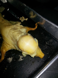
Before we cut open the pig, we needed to determine the sex of the fetal pig. We could not find any opening near the umbilical cord, so we thought it might be female. However we thought there was a possibility that if this specimen was male, its opening near the umbilical cord could be undeveloped yet, since it was so small compared to the specimen other groups had.

Then we started to cut the skin of the abdominal area. We found out that there was a fat layer between the skin and the internal organs. This proved that lipids were great protection.

Finally we cut open the abdomen of the pig. There was a lot of grayish liquid pouring out. It could not have been blood since it must have been drained out already. It might be substances that preserved the specimen.

I was surprised to see that the liver was so large in terms of the size. It looked like it was 3 times larger than the intestines. Then I remembered that this was a fetal pig, which meant there wasn't anything in its intestines, so the intestines were flat and small in size. Additionally, we saw two red blood vessels at the lower abdominal cavity. We inferred they were renal arteries, and they were dyed red for students to see better.

Removal of the organs. We needed to be very careful in order not to damage other organs.

Outcomes of day 1! We had isolated liver, stomach, pancreas,kidneys, small and large intestines.
The pancreas was not easy to find. It was a leaflet-like structure that was close to the stomach, and was easy to be damaged.

After 1 day, the specimen had become very dehydrated. We started to open its upper body. The first organs exposed were the lungs.The heart was hidden between the lungs. After removing the lungs and the heart, we tried to locate the thyroid glands, which should have been near to the trachea. However, we were not sure if we had found it, or some adipose tissue.

Moving up to the brain. Since the specimen was very undeveloped, its skull was not bone yet but cartilage, which was quite soft but strong enough to hold the brain within. Due to its fragility, we cannot use scissors or knife. Instead, we used a very sharp needle-like tool to gently divide the head into four areas (the white lines on the head in the photos below), then we used small tweezers to open up its skull. The process took a lot of time and required concentration and refined movement.
After that, we tried to isolate the brain and the brain stem attached. We cut open the spine at the back of the head a bit.
Like all the other groups, we had failed to isolate the brain stem attaching to the brain.
Out of curiosity, we wanted to know what the brain felt like, and we all touched it. The result was that it felt like jelly.


That was the end of the of this dissection. Here is an overview of all the organs we isolated.

No comments:
Post a Comment